Key Takeaways
- Operative hysteroscopy allows clinicians to evaluate and treat selected conditions within the uterine cavity.
- Preparation, visualization, fluid monitoring, and communication are central parts of procedural safety.
- Patient history, suspected pathology, medical needs, and fertility goals should shape the treatment plan.
- Not every cause of abnormal uterine bleeding can be diagnosed or treated with hysteroscopy.
Operative hysteroscopy uses a hysteroscope, a narrow camera-based instrument passed through the vagina and cervix, to provide a view inside the uterus. Depending on the clinical situation, the procedure may be diagnostic, therapeutic, or both.
For patients with abnormal uterine bleeding or a suspected finding inside the uterine cavity, hysteroscopy can help a clinician directly assess the location and appearance of tissue. It may also allow selected polyps, fibroids, adhesions, or retained tissue to be addressed during the same procedure when clinically appropriate.
Why Operative Hysteroscopy Still Matters
Hysteroscopy is commonly used to investigate heavy menstrual bleeding, irregular bleeding, suspected endometrial polyps, and submucosal fibroids. [1] It may also be used to obtain a tissue sample for pathology or to evaluate certain intrauterine abnormalities. The American College of Obstetricians and Gynecologists notes that hysteroscopy may be performed in an office, hospital, or ambulatory surgery setting, depending on the planned procedure, patient needs, and anesthesia requirements. ACOG's guidance on hysteroscopy provides an overview of common uses, preparation, recovery, and potential risks. [2]
Imaging studies, such as ultrasound or saline infusion sonography, can identify potential abnormalities, but they do not always provide a complete picture of the uterine cavity. Direct visualization may help confirm whether a finding is focal, where it is located, and whether tissue removal or sampling should be considered. [3]
Common Intrauterine Findings
Endometrial Polyps
Endometrial polyps are growths that arise from the lining of the uterus and may be associated with spotting, irregular bleeding, or heavy bleeding. When a polyp is identified within the cavity, hysteroscopy can help a clinician visualize and remove it in a targeted manner. For example, a patient with recurring spotting may first undergo imaging, then proceed to hysteroscopy if the results suggest a focal intracavitary lesion.
Submucosal Fibroids
Submucosal fibroids extend toward or into the uterine cavity and can contribute to heavy bleeding and, in some situations, fertility concerns. Their management depends on size, location, depth of penetration into the uterine wall, symptoms, overall health, and the patient's reproductive goals. A hysteroscopic approach may be appropriate for selected fibroids, but not all fibroids can be treated this way. [4]
Other Findings
Other conditions that may be evaluated or managed hysteroscopically include intrauterine adhesions, retained pregnancy tissue, abnormal-appearing endometrial tissue, and selected congenital uterine findings. Hysteroscopy is not a treatment for every gynecologic condition, and it is not a treatment for endometriosis or uterine cancer. When tissue is removed or sampled, a pathology review may be needed to guide the next clinical step.
Planning Before the Procedure
Thoughtful planning begins with a clear understanding of why the procedure is being performed and what outcome is expected. The pre-procedure discussion may include:
- The suspected cause of symptoms and the planned procedural goal.
- Pregnancy testing when appropriate.
- Medication, allergy, bleeding risk, and prior procedure review.
- Anesthesia and pain-management options.
- Fertility goals and the importance of uterine preservation.
- Whether tissue may be collected for pathology.
- Expected recovery, follow-up timing, and symptoms that require prompt medical attention.
Building a Clear Procedural Workflow
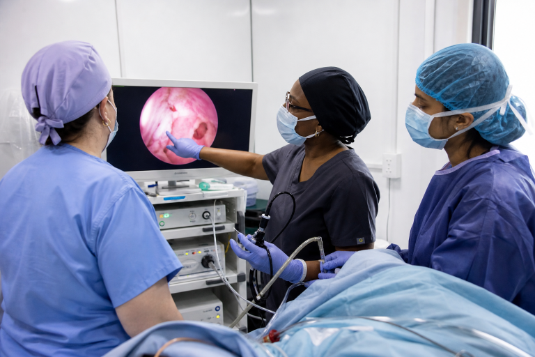
Before the procedure begins, the clinical team should align on the plan, anticipated pathology, and available equipment. A practical setup check can include the hysteroscopic imaging components, light source, distention-fluid system, suction, collection method, energy equipment when indicated, and backup supplies. A formal time-out should confirm the patient, planned procedure, consent, allergies, and relevant safety considerations.
Clear role assignment also matters. The surgeon, nursing staff, anesthesia team, and other support personnel should know who is tracking fluid, who is documenting key events, and how the team will communicate if the procedure changes. Standardized preparation can reduce uncertainty and help avoid unnecessary interruptions.
Visibility and Fluid Management
Maintaining a stable view of the uterine cavity is essential during diagnostic assessment and tissue removal. Blood, bubbles, tissue fragments, poor positioning, and inadequate fluid exchange can reduce visibility. When the view becomes limited, the team should pause, identify the cause, and restore visualization before proceeding.
Distention fluid opens the uterine cavity so the clinician can see and work within it. Fluid deficit is the difference between the volume introduced and the volume recovered. Excessive absorption can lead to serious complications, so fluid input, output, pressure, and deficit should be monitored throughout the procedure. Professional guidance recommends designating a team member to regularly measure and report fluid status, with lower thresholds for patients with relevant comorbidities or limited access to acute care resources. AAGL fluid-management guidance emphasizes selecting an appropriate distention medium, minimizing absorption, and recognizing excessive fluid deficit early.
Choosing a Tissue-Removal Method
Mechanical, electrosurgical, and other hysteroscopic tissue-removal approaches each have different considerations. The appropriate method depends on the lesion's size, type, and location, as well as the planned clinical objective, available equipment, and clinician experience. Decisions should account for visualization, controlled tissue removal, specimen recovery, fluid management, and patient-specific needs rather than procedure time alone.
Safety Beyond the Procedure
Discharge instructions should explain what recovery may look like and when to seek care. Mild cramping or light bleeding can occur after hysteroscopy, but patients should contact their clinician promptly for heavy bleeding, fever, chills, severe or worsening pain, fainting, shortness of breath, or foul-smelling discharge. The follow-up should also clarify when the pathology results are expected and who will discuss them.
Questions Patients Can Ask
- What is the suspected cause of my bleeding or symptoms?
- Will this procedure be diagnostic, therapeutic, or both?
- Will tissue be removed or sent for pathology?
- What are the alternatives to hysteroscopy?
- Could this procedure affect future fertility?
- How will fluid levels be monitored during the procedure?
- What symptoms should prompt an urgent call after discharge?
Better Hysteroscopy Starts With Better Preparation
Effective operative hysteroscopy depends on patient selection, a clear procedural plan, reliable visualization, careful fluid tracking, and coordinated teamwork. Honest conversations about expected findings, alternatives, recovery, and follow-up help patients participate meaningfully in their care. Individual evaluation by a qualified clinician remains essential because symptoms, medical history, and treatment goals vary from person to person.
References
1. Cleveland Clinic. Hysteroscopy. https://my.clevelandclinic.org/health/treatments/10142-hysteroscopy
2. American College of Obstetricians and Gynecologists. The Use of Hysteroscopy for the Diagnosis and Treatment of Intrauterine Pathology. https://www.acog.org/clinical/clinical-guidance/committee-opinion/articles/2020/03/the-use-of-hysteroscopy-for-the-diagnosis-and-treatment-of-intrauterine-pathology
3. Johns Hopkins Medicine. Sonohysterography. https://www.hopkinsmedicine.org/health/treatment-tests-and-therapies/sonohysterography
4. National Library of Medicine. Management of uterine fibroids in the patient pursuing assisted reproductive technologies. https://pmc.ncbi.nlm.nih.gov/articles/PMC3444289/

Leave a Reply